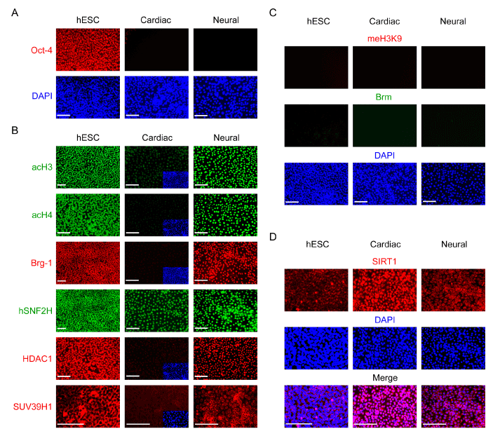
(A) NAM (cardiac) or RA (neural) treatment induced differentiation under the defined culture system, as indicated by the appearance of Oct-4 (red) negative cells within the colony. Untreated cells were used as the control.
(B) The strong expression and nuclear localization of a set of active chromatin modifiers in untreated pluripotent hESCs (control), including acetylated histones H3 (acH3, green), acetylated histones H4 (acH4, green), ATP-dependent active chromatin-remodeling factors Brg-1 (red) and hSNF2H (green), and HDAC1 (red), and cytoplasmic localization of histone methyltransferase SUV39H1 (red). NAM-induced cardiomesodermal cells (cardiac) displayed significantly decreased levels of histone H3 and H4 acetylation, and Brg-1 and HDAC1 expression, while RA-induced neuroectodermal cells (neural) retained the similar expression levels and localization patterns. All cells are indicated by DAPI staining of their nuclei (blue) in the insets.
(C) The weak or undetectable expression of repressive chromatin remodeling factor Brm (green) and K9 methylated histone H3 (red).
(D) NAM induces nuclear translocation of NAD-dependent HDAC SIRT1 (red). All cells are shown by DAPI staining (blue) of their nuclei. Scale bars: 10 Ám.