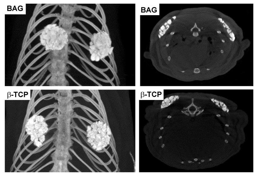
 The figure shows representative images for implanted BAG and ß-TCP
granules at week 8. Dorsoventral and transversal views on computed
tomographic imaging confirmed standardized surgical procedures at week 4
and 8, since no volume differences were measured between surgical sites.
Furthermore, biomaterials remained at the original implantation sites over
time.