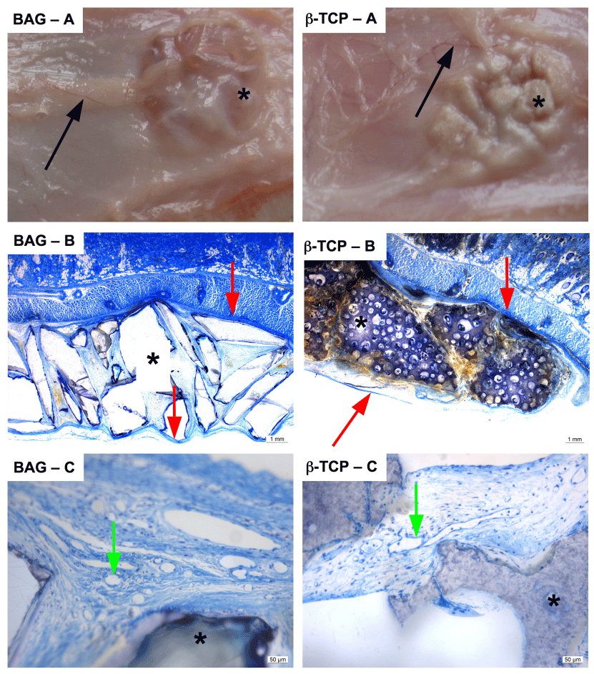
 The figure shows representative images for implanted BAG and ß-TCP granules
at week 8. At weeks 4 and 8, samples were harvested and demonstrated similar
macroscopical appearance independent on biomaterial implanted. Vessels
(BAG/ß-TCP – A; black arrows) were sprouting centripetally into all capsules
indicating viability. A translucent and thin capsule (BAG/ß-TCP – B; red arrows)
formed around all specimens. Infiltrating vessels provided vascularization of
periimplantar granulation tissue (BAG/ß-TCP – C; green arrows). Asterisks
indicate biomaterials.