 The figure shows representative images for BAG and β-TCP at week 8. BAG
– A = healthy, vascularized granulation tissue was formed around all granules
containing spindle-shaped cells (red arrow); BAG – B = macrophages (green
arrow) and foreign body giant cells (red arrow) were simultaneously observed;
β-TCP – A = typical appearance of foreign body giant cells (red arrow)
accumulating at the surface of a β-TCP granule; β-TCP – B = preosteoblastic
cell nests (green arrow) were only observed after seeding with hASCs and/
or BMP-2 supplementation, but early calcification along β-TCP granules
(red arrow), was only observed after hASC seeding. Asterisks represent
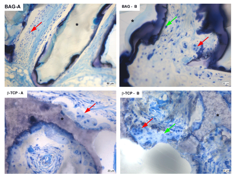
biomaterials. The figure shows representative images for BAG and β-TCP at week 8. BAG
– A = healthy, vascularized granulation tissue was formed around all granules
containing spindle-shaped cells (red arrow); BAG – B = macrophages (green
arrow) and foreign body giant cells (red arrow) were simultaneously observed;
β-TCP – A = typical appearance of foreign body giant cells (red arrow)
accumulating at the surface of a β-TCP granule; β-TCP – B = preosteoblastic
cell nests (green arrow) were only observed after seeding with hASCs and/
or BMP-2 supplementation, but early calcification along β-TCP granules
(red arrow), was only observed after hASC seeding. Asterisks represent
biomaterials. |