|
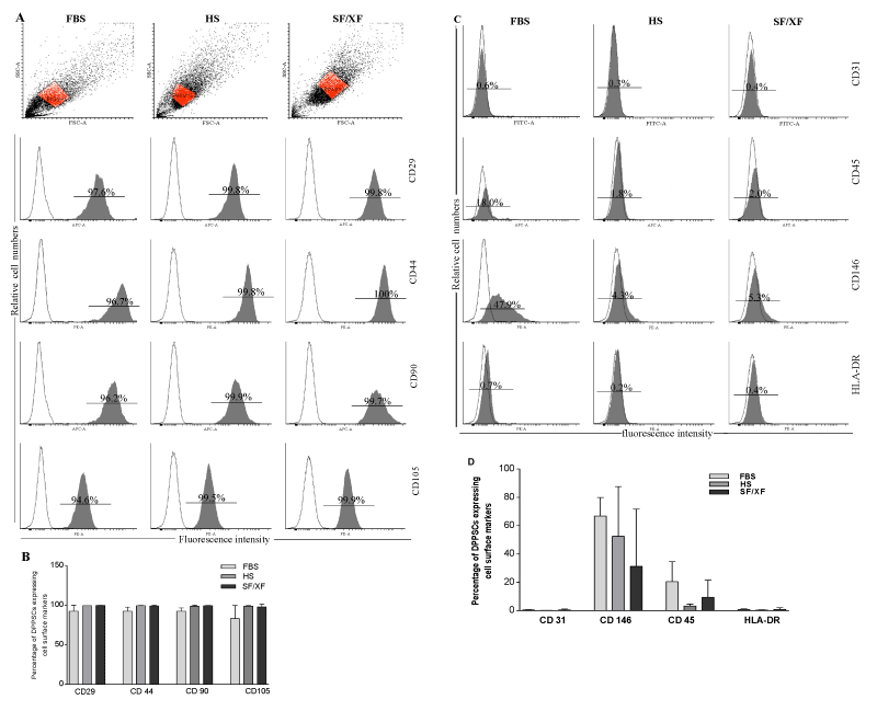
| Figure 4: Surface marker expression of DPSCs cultured in FBS-M, HS-M and SF/XF-M was analyzed by flow cytometry. (A,C) Histograms demonstrating the forward and side scatter, relative cell count (y-axis) and fluorescence intensity (x-axis), with unstained control cells (empty histogram) and cells stained with antibodies against the surface proteins (filled histogram). (B,D) Column graph of flow cytometry data of surface marker expression levels, the bars represent the mean ± SD (n = 4). DPSCs: dental pulp stem cells; FBS-M: fetal bovine serum medium; HS-M: human serum medium; SF/XF-M: serumfree/xenofree medium. |