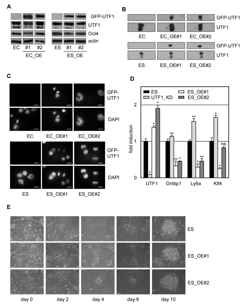
A) Western analysis of wild type P19CL6 (EC) and IB10 (ES) cells and GFPUTF1 expressing P19CL6 (EC_OE#1 and #2) and IB10 (ES_OE#1 and #2) cells. Cell lysates were analyzed with the antibodies indicated, UTF1 and GFP-UTF1 were both detected with an αUTF1 antibody on the same blot.
B) Subnuclear fractionation of ES and EC cells. Fractions were immunostained with an UTF1 antibody. Fractionation abbreviations: F, free diffusing/ cytoplasmic fraction; D, DNaseI released fraction; AS, ammonium sulfate fraction; HS, high salt fraction; M, nuclear matrix fraction.
C) Deconvoluted fluorescent images of wild type (ES and EC) and GFP-UTF1 expressing (EC_OE#1, EC_OE#2, ES_OE#1 and ES_OE#2) ES and EC cell lines. Cells were counterstained with DAPI.
D) Effect of GFP-UTF1 expression on UTF1 target genes. Transcript levels of UTF1 and the indicated UTF1 target genes were determined using quantitative RT-PCR using GAPDH as an internal standard. Mean expression levels and the standard deviations are depicted; *: p<0.01; **: p<0.05 and ns: not significant.
E) Phase contrast images of wild type (ES) and GFP-UTF1 expressing (ES_ OE_#1 and #2) IB10 ES cells 2-10 days after LIF withdrawal.