 A) Phase-contrast images of DMSO-treated (0, 2, 5 and 10 days) wild
type (ES) and GFP-UTF1 overexpressing ES cells (ES_OE#1 and #2).
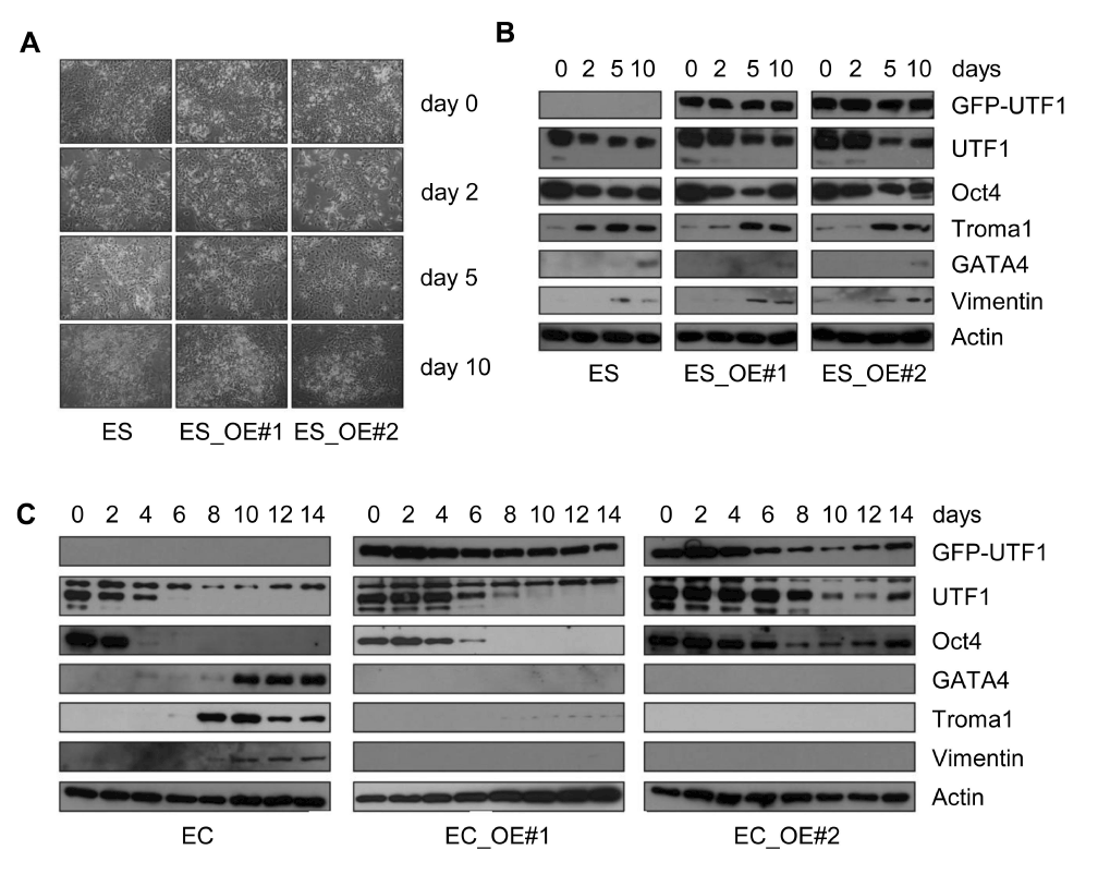
A) Phase-contrast images of DMSO-treated (0, 2, 5 and 10 days) wild
type (ES) and GFP-UTF1 overexpressing ES cells (ES_OE#1 and #2).
B) Western blots of DMSO-induced differentiation (0, 2, 5 and 10 days) of wild type (ES) and GFP-UTF1 overexpressing ES cells (ES_OE#1 and #2). Protein lysates were analyzed with antibodies against GFP, UTF1, Oct4, Troma1 (endoderm), GATA4 (endoderm and mesoderm), and Vimentin (mesoderm). Actin staining was performed as a loading control.
C) Western blots of DMSO-induced differentiation of wild type (EC) and GFP-UTF1 overexpressing P19CL6 EC cell lines (EC_OE#1 and #2). Protein lysates were collected at the time points indicated and analyzed with antibodies against GFP, UTF1, Oct4, GATA4, Troma1 and Vimentin. Actin staining was performed as a loading control.