 A) Phase-contrast images of RA-treated (0, 2, 5 and 10 days) of wild
type (ES) and GFP-UTF1 overexpressing ES cells (ES_OE#1 and #2).
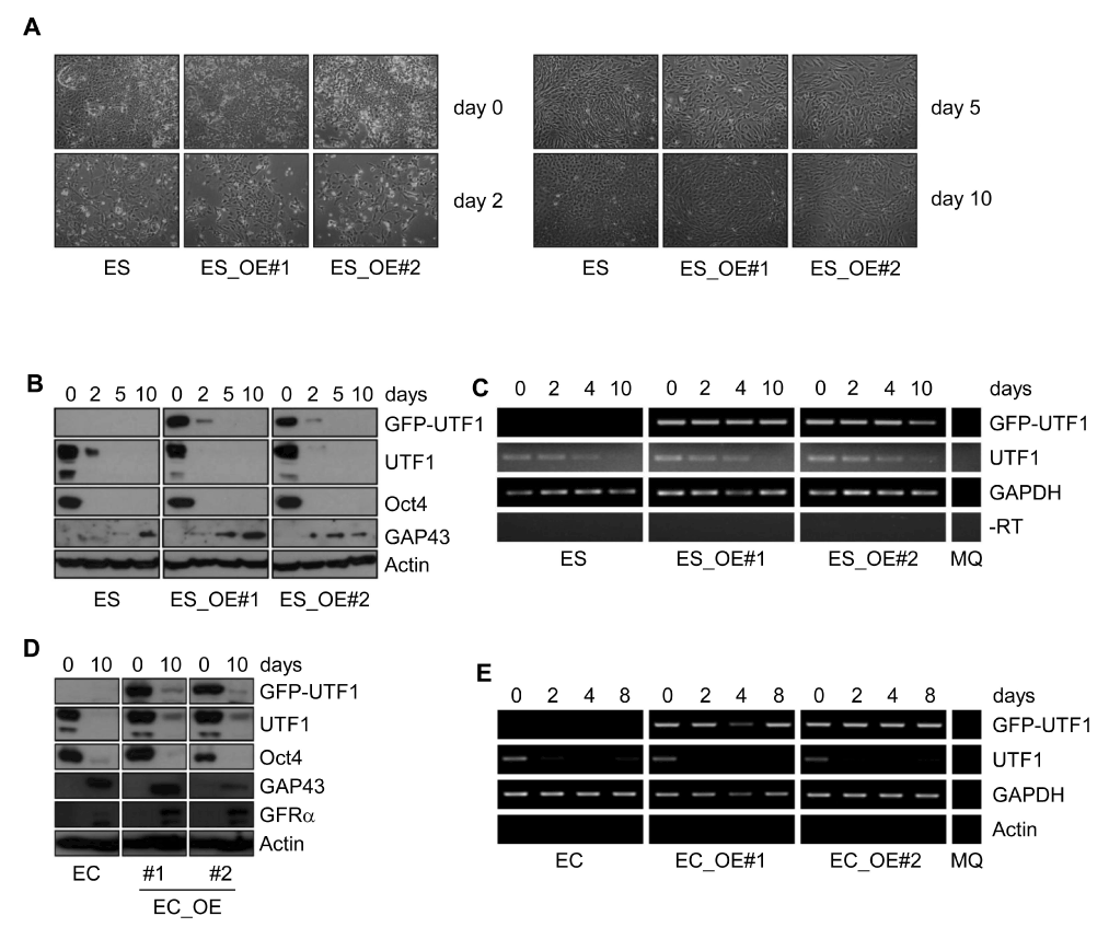
A) Phase-contrast images of RA-treated (0, 2, 5 and 10 days) of wild
type (ES) and GFP-UTF1 overexpressing ES cells (ES_OE#1 and #2).B and D) Western blots of RA-induced differentiation (0, 2, 5 and 10 days) of wild type (ES and EC) and GFP-UTF1 overexpressing IB10 ES and P19CL6 EC cells (OE#1 and #2). Protein lysates were analyzed with antibodies against GFP, UTF1, Oct4, GAP43 (ectoderm) and GFRα (ectoderm). Actin staining was performed as a loading control.
C and E) RT-PCR analysis of UTF1 and GFP-UTF1 expression in wild type (ES and EC) and GFP-UTF1 overexpressing IB10 ES and P19CL6 EC cells (OE#1 and #2) during RA treatment. Glyceraldehyde-3-phosphate dehydrogenase (GAPDH) expression was used as a control. In the -RT lanes, reverse transcriptase was omitted from the reverse transcriptase reactions to control for genomic DNA contamination and amplified using GAPDH primers. In the MQ lane, no template was added to the PCR reactions.