|
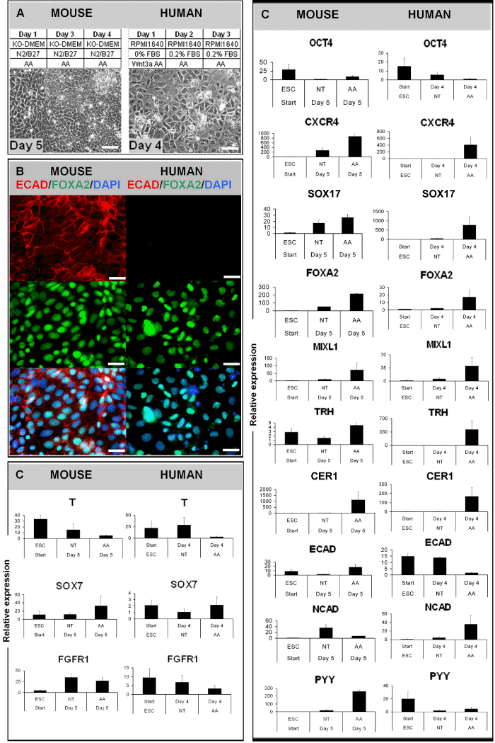
| Figure 3: Analysis of putative human and mouse definitive endoderm prior to grafting. (A) The differentiation protocols for deriving putative mouse and human DE from ESCs and bright-field images of mouse and human DE. Scale bars: 25 μm. (B) Immunofluorescence stainings (Sox17 and Ecad/FoxA2) of human and mouse DE. Nuclei are indicated by DAPI staining. Scale bars: 50 μm. (C) Relative mRNA-expression of genes in undifferentiated cells (ESC) as well as differentiated progeny treated with or without Activin A. NT = no treatment, AA = Activin A treatment. |