|
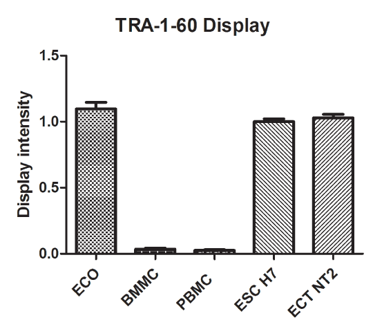
| Figure 6: Quantified ECO cell surface display of TRA-1-60. The pixel intensity values were measured on the TRA-1-60 scFv labeled immunoblots of the cell lysates as described in the figure 5. Labels: ECO – patients’ samples (n=6); PBMC – peripheral blood mononuclear cells (n=6); BMMC – bone marrow mononuclear cells (n=6); ESC – cultures of the human embryonic stem cells H1, H7, H9 (n=3); ECT -cultures of the human testicular embryonal carcinoma cells NT2D1 (n=1). Statistical significance accepted at the p < 0.001. The samples were run and quantified in triplicates. |