|
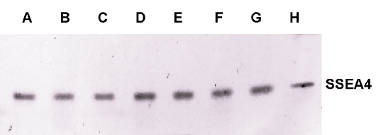
| Figure 7: SSEA-4+ display on the ECO cells. The ECO cells were disintegrated, electrophoresed, transferred onto the PVDF membranes, and labeled with the Fvs targeting SSEA-4. Labels: ECO – patients’ samples (encoded A-F); ESC – culture of the human embryonic stem cells H7 (G); ECT -culture of the human testicular embryonal carcinoma cells NT2D1 (H); PBMC – peripheral blood mononuclear cells; BMMC – bone marrow mononuclear cells (empty lanes not shown). The samples were run and quantified in triplicates. All the immunoblots were quantified as presented in the figure 8. |