|
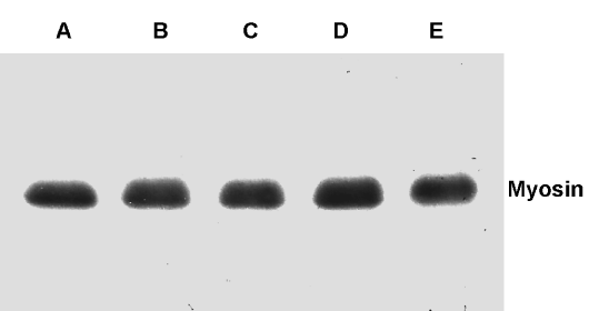
| Figure 15: Differentiation of the ECO cells into muscle. The ECO cells were induced to differentiate into the cardiac muscles. The cells were homogenized, electrophoresed, and transferred onto the PVDF membranes. The transfers on the membranes were labeled with the antibodies against cardiac muscle myosin. Labels: ECO – patients’ samples (encoded A-C); cardiac muscle myosin (D); human cardiac muscle (E); PBMC – peripheral blood mononuclear cells; BMMC – bone marrow mononuclear cells (no labeling - empty lanes not shown). |