 |
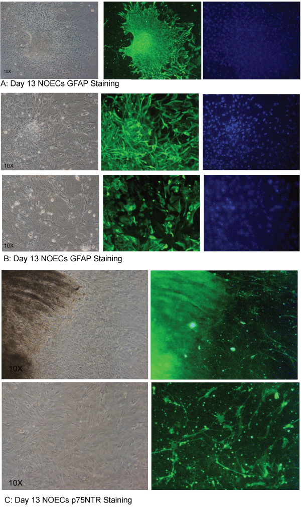
| Figure 2: The confluent flask was immuno-cytochemically characterized on day 13 through Glial Fibrillary Acidic Protein (GFAP) from 2-day-old Neonate of Sprague–Dawley strain. Microscopic examination (Nikon Eclipse Ti Phase Contrast Microscope) suggested dense population of heterogeneous cells through spatial juxta-positioning of unstained and immunostained areas through GFAP (A). Moreover, both spindle shaped astrocyte like cells and the flattened sheet like Schwann cells were also observed through GFAP staining (B). However, FITC labelled p75 NTR Protein stained cells on day 13 specifically stained only spindle-shape astrocyte like cells (C). The cells were photographed on magnification 10X. |