|
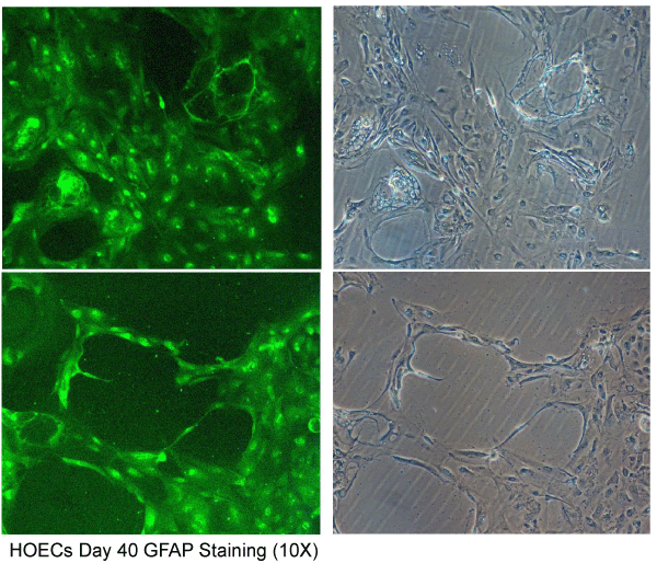
| Figure 9: By 6 weeks the cultured cell mass demonstrated degenerative changes where the cell morphology appeared dissoluting established through FITC labelled GFAP. This is in agreement with the unstained phase contrast microscopy pictures observed at 10X. |