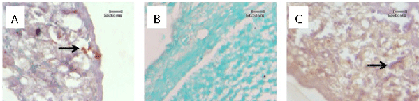
Figure 1:
Immunohistochemical staining for GDF-9 in rat ovarium of (A) control, (B) cisplatin and (C) cisplatin+BMT groups show the immunoreactive cells with brown colour.