|
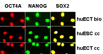
| Figure 7: The stem cells biopsied directly from the primary tumors of the patients diagnosed with the embryonal carcinoma of the testes (the patients encoded 001-009), the cultured human embryonic stem cell lines (H1, H13, H14) (huESC cc), and the cultured cells from metastasis to lungs of the testicular embryonal carcinoma (NT2D1) (huECT cc) were heavily saponinpermeabilized and triple-labeled with the Fvs targeting OCT4A - red, NANOG - green, and SOX2 – yellow DNA (violet). Specificity and sensitivity of the Fvs were validated on the blots as shown in the figure 6. All the batches for every patient were labeled in triplicates. From each batch, the images of ten randomly selected fields of view were acquired via the three acquisition channels without correcting the wavelength induced shift. The images were acquired with multiphoton excitation fluorescence. The images presented in the figure were acquired from the primary tumors of the patient (huECT bio) (Patient encoded 001), the cultured human embryonic stem cell line – cell line H1 (huESC cc), and the cultured cells from metastasis to lungs of the testicular embryonal carcinoma – cell line NT2D1 (huECT cc). The images presented in the figure are representative to all the patients and the controls studied. The HFW: 200μm. |