|
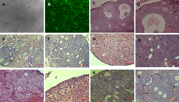
| Figure 2: Transplantation of GFP-transfected human amniotic epithelial cells (hAECs) into chemotherapy-sterilized recipient mice. (A) hAECs cells grown to 85% density. (B) GFP-transfected hAECs. Representative H&E micrographs of ovary sections from: non-sterilized normal control mice (C, D); sterilized non-transplanted mice after a 7 day-recovery period (E), a 14 day-recovery period (F), a 21 day-recovery period (G) and a 2 month-recovery period (H ) showing stroma, and atretic primordial or primary follicles; sterilized recipient mice 7 days (I), 14 days (J), 21 days (K) and 2 months (L) following transplantation of hAECs. No obvious follicles were shown in recipients 7 days following hAECs transplantation (I), however, the cavities reduced obviously compared with (E). Primordial follicles were visible in J, primary follicles were visible in K and large antral follicles were shown in L. Arrows indicated follicles at various stages of maturational development. Scale bars=100 μm. |