 |
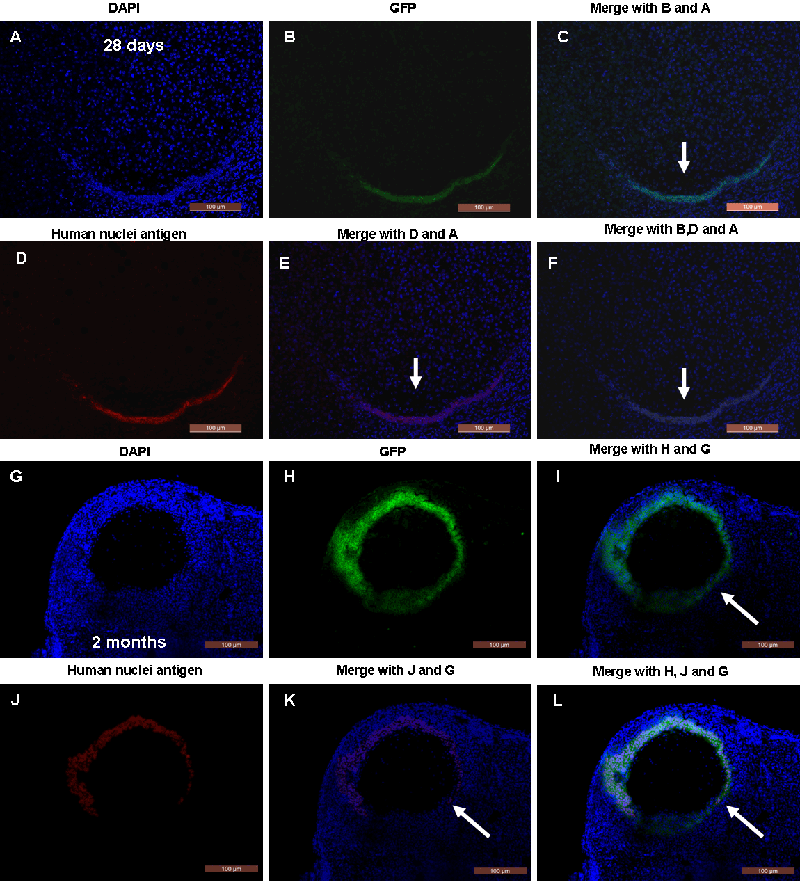
| Figure 4: Double-staining with GFP and human nuclei antigen were performed to look for the derivation of GFP positive cells in recipient ovaries after hAECs transplantation. (A-F) Ovarian sections stained with GFP and anti–human nuclei antibody revealing grafted hAECs 28 days following hAECs transplantation. (G-L) GFP staining was co-locolized with human nuclei antigen in antral follicles of recipient ovaries 2 months following hAECs transplantation. Arrows indicated the doublestaining pattern in ovaries. Scale bars, 200 μm (G-L), 100 μm (A-F). |