|
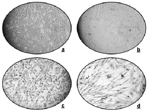
| Figure 1: Representative images of peripheral blood (PB)-derived mesenchymal stem cells (MSCs): light microscopy at 20x (a) and 40x (b) magnification and after staining with haematoxylin at 20x (c) and 40x (d) magnification. Scale bars represent 50μm. |