|
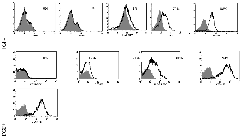
| Figure 3: Expression of surface markers by patient MSCs expanded until confluence with or without FGFb. Cells cultured with FGFb were investigated on day 16, without FGFb – on day 21. The percent of cells in the positive gate is indicated on the histogram. Samples are presented in the shaded plots, corresponding isotype controls are shown as open lines. |