|
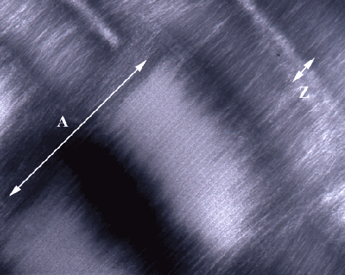
| Figure 2: The molecular architecture of the human cardiac muscle is revealed by electron energy loss spectroscopy (EELS). The fresh cardiac muscle was cryoimmobilized and cryosectioned followed by embedding / negative staining. The image was acquired at the zero energy loss, with the contrast tuned setting of the energy filter at the accelerating voltage 300keV. All the samples were prepared in triplicates. This image is representative for all the samples tested. The Z-line (Z) is the part of the sarcomeres in which actin filaments bind to its α-actinin. The A-band is formed by thick myosin filaments. It also includes the cross-bridges between myosin heads and thin actin filaments; the H-zone is a bare portion of thick myosin filaments; the M line in the H-zone’s center. I-band, consisting of the thin, actin filaments, is the part of the sarcomere between the Z-line and A-band. The densely packed biomolecular clusters of Z-line and A-band exclude the molecules of the negative stain; hence they appear with light contrast. The H-zone and I-band contain more space between filaments, which absorb and retain a lot of contrast, therefore appear as the very dark structures. HFW: 3750 nm. |