 |
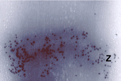
| Figure 3: The localization of α-actinin in the Z-line is revealed by electron energy loss spectroscopy (EELS). The fresh cardiac myofibrils were stained with the bioengineered, monospecific tetravalent antibodies anti-α-actinin covalently linked to the core-shell nanoparticles. The image was acquired at the zero energy loss, with the contrast tuned setting of the energy filter at the 300keV accelerating voltage. The Z-line’s α-actinin is heavily labeled (black dots), while the background is label-free. Vanadate-PVP based negative stain / embedment offered lower contrast of the imaged cardiac muscle myofibril, but promoted a better contrast for the nanoparticles. The threshold driven nanoparticles’ counts facilitated determination of specificity of the labeling, which was accepted at the statistical significance p < 0.001. The high specificity of this and other Fvs was required for bioengineering of the highly specific htAbs with no cross-reactivity. All the samples were measured in triplicates. This image is representative for all of the samples and all of the Fvs tested. HFW: 500 nm. |