|
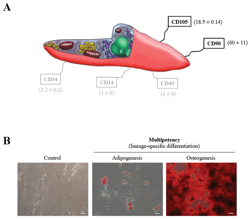
| Figure 1: Characterization of primary cultures of human UCBMSCs. A) Schematic illustration of an UCBMSC showing fluorescence intensity data in arbritary units from surface antigen expression analysis by flow cytometry. UCBMSCs were strongly positive for CD105 and CD90 (60 ± 11), and consistently negative for CD45, CD34, and CD14. Values are expressed as mean ± SD. B) Standard MSC differentiation into adipogenic and osteogenic cell-lineages. Images display a control, nondifferentiated (left) and differentiated cell cultures following staining with Oil red O (middle) and Alizarin red S (right). Scale bars: 100 μm. |