|
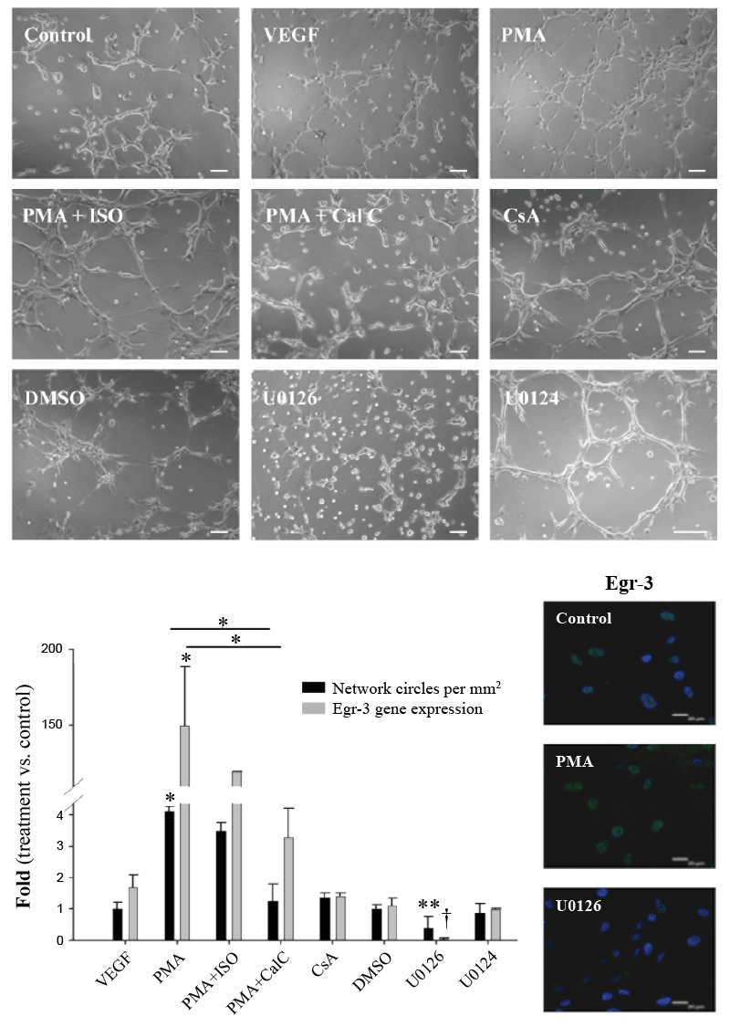
| Figure 2: Modulation of Egr-3 expression levels and angiogenic capacity by UCBMSCs. Representative images and quantification of the cell network-forming capacity, measured as the number of network circles per unit area, of UCBMSCs after 2 h in Matrigel following treatment with a specific agonist (PMA) and inhibitors (Cal C and U0126) of PKCα/MEK/Egr-3 signaling. Scale bars: 100 μm. Histogram represents the relative levels of Egr-3 gene expression and cell network formation compared with those in control or untreated cells. DMSO and U0124 were used as vehicle and negative control, respectively. Note that there is equivalence in the scales of gene expression and network circles/mm2, maybe indicating the close relationship between these two cellular events. Data are from three independent experiments and all values are expressed as mean ± SD. Specific detection of Egr-3 protein in control, PMA- and U0126-treated UCBMSC cultures by indirect immunofluorescence. *P<0.001, **P=0.016 and †P=0.015. Scale bars: 20 μm. A minimum of 10 microscopic fields per condition and experiment (n=3) were analyzed with similar results. |