 |
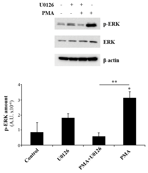
| Figure 4: PMA induces phosphorylation of ERK1/2 in UCBMSCs. Western blot panels show that ERK1 (44 kDa) and ERK2 (42 kDa) were phosphorylated following exposure to PMA. In contrast, addition of U0126 suppressed PMA-mediated ERK1/2 phosphorylation. Histogram depicts protein levels as arbitrary units (A.U.), which were determined by densitometry in three independent experiments. A 3.6-fold activation of ERK1/2 was detected in PMA-treated relative to control cells. N=3 for each experimental condition with similar results. Values are expressed as mean ± SD. *P=0.021 and **P=0.014. |