|
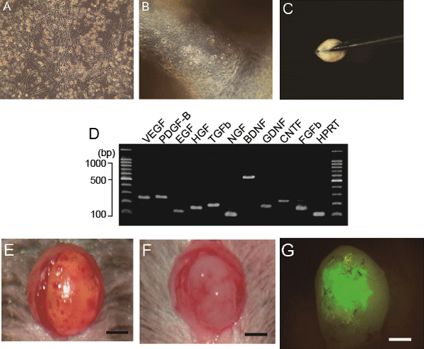
| Figure 2: Preparation of sheet-pellets and cell suspension, expression of neurotrophic and vascular growth factors, and macroscopic observations of damaged and transplanted bladder. A and B: Cultured cell behavior just before and after the detachment. C: A gel-like sheet-pellets being picked up with forceps. D: RT-PCR for sheet-pellets. E: State of surface delamination. F: Pasted sheet-pellets. G: Engraftment of GFP-positive tissues at 4 wk after transplantation. Used primers were presented in Table 1. Bars=5.0mm D: Expressions of neurotrophic and vascular growth factors in sheet-pellets. |