 |
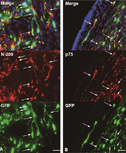
| Figure 6: Contribution of donor-derived GFP+ cells to reconstitution of nervous networks. Note that the close relationship between GFP+ cells and N-200+ nerve axons (arrows in A) are observed. Similarly, co-staining of GFP+ and p75+ cells were also evident (arrows in B) Bars=50μm. |