|
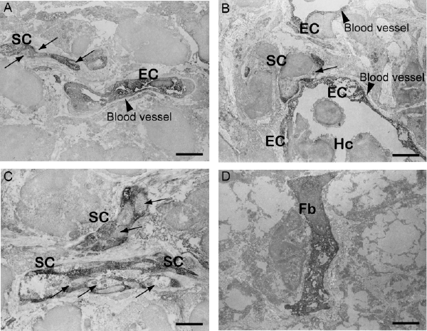
| Figure 7: Immunoelectron microscopic detection of engrafted GFP+ cells. Transplanted donor-derived GFP+ cells were stained with anti-GFP antibody and visualized by DAB (dark dots). Differentiation of transplanted donor cells into Schwann cells (arrows in A-C), endothelial cells (arrowheads in A and B), and fibroblasts (D) were clearly evident. SC, Schwann cell; EC, endothelial cell; Hc, hematocyte; Fb, fibroblast. Bars=2μm. |