 |
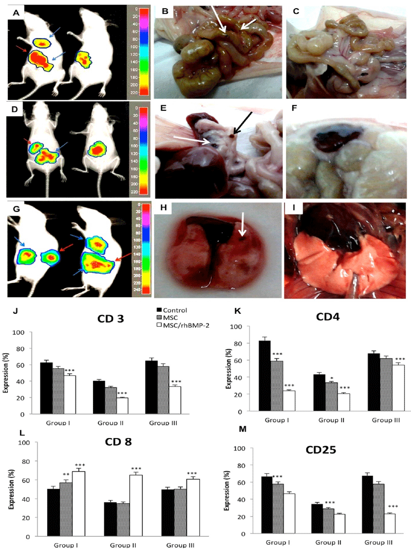
| Figure 3: Expression lymphocytes and metastasis markers. Tumoral development was detected by bioluminescence (A, D, and G, arrows) and necropsy in the control animals of the group III. In: (B, E, and H): metastasis in intestine, spleen and lung, respectively in animals from group III (arrows). In (C, F, and I) no metastasis was observed in the same organs in animals from group III with association of MSC/rhBMP-2. In (J, K, and M) was observed a decreased in the expression of CD3, CD4, and CD25, respectively by flow cytometry in the analyzed groups. (L): In the groups with the association of MSC/rhBMP-2 there was an increased in the expression of CD8. Statistical differences were obtained by analysis of variance (*** p < 0.001, ** p < 0.01, and * p< 0, 05). |