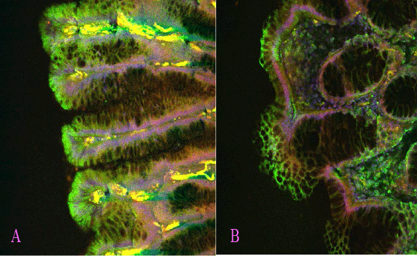
Showing a comparison between a near normal section of villi (A) and an abnormally developing villi (B) from the same patient. The abnormal polyp tissue (B) exhibits a con-commitant loss of CKI protein expression due to the loss of cytoplasmic APC in growing cancerous cells. Note the transition of CKI protein expression to a patchy discontinuous appearance in the basal cell-membranes of the growing polyps in panel B, in contrast to a continuous basal membrane and cytoplasmic localization of the protein while APC is still cytoplasmic (arrow in A). The arrow in panel B suggests loss of this cytoplasmic component of APC in the mutant cells within a mixed population of normal and cancerous tissues.