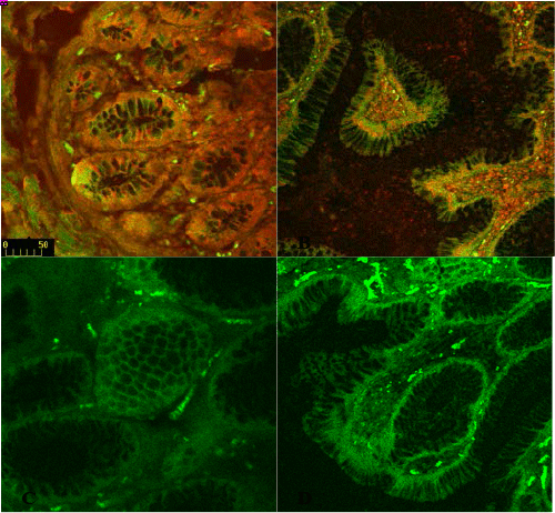
The protein localization of APC and hDlg proteins in (A) normal colonic crypts and (B) polyp tissue. Note the normal basolateral membrane localization of hDlg in the polyp structure (B) (arrowhead) which is also evident in the normal crypts (A). Panel (C) shows an abnormal crypt (arrow) exhibiting loss of apicobasal polarity and the panel (D) depicts abnormally developing villi from the colon of this tubulovillar case giving rise to early polyps. Note that hDlg is mislocalized due to the architectural disturbances in the dysplastic cells of crypts and villi in these premalignant tissues, which lose their apico-basal polarity and not due to the loss or degradation of APC protein per se ( B), which tends to accumulate in the lumen of the gut and crypts (red arrows). Figures A and C are acquired at 35X whereas; Figures B and D are at 20X.