|
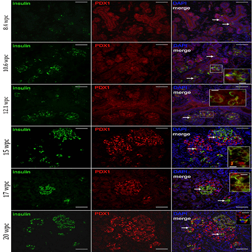
| Figure 4: Insulin (green) and PDX1 (red) expression during the first and second trimester of human fetal development (1- 24 wpc), as demonstrated by samples 8.4, 10.6, 12.1, 15, 17, and 20 wpc. Arrows demonstrate examples of co-expressing insulin and PDX1 cells. In the first trimester, PDX1 expressing cells are located in numerous duct-like structures, and insulin expressing cells co-express PDX1. At 8.4 wpc, scattered insulin expressing cells are located amongst the duct-like PDX1 expressing cells. As more insulin expressing cells aggregate at 10.6 and 12.1 wpc, the islet-like structures seem to bud off from the PDX1 expressing duct-like structures. In the second trimester, PDX1 expression is localized mainly to islet structures, and PDX1 expression is most pronounced in cells surrounding the insulin expressing cells. All insulin expressing cells co-express PDX1. The islet structures have less spatial association to the duct-like PDX1 expressing structures. DAPI nuclear counterstain (blue). Areas denoted by a box are shown at higher magnificiation (inset). Higher magnification images of 12.1, 15 and 17 wpc are shown without DAPI. Scale bars are 50 and 10 μM respectively. |