|
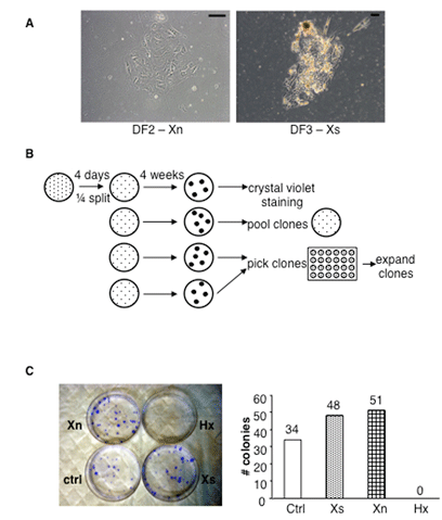
| Figure 2: The SACK agents Xn and Xs increase the efficiency human pancreatic cell strain expansion. A. Phase micrograph examples of primary cell colonies obtained after 4 days of culture of pancreatic cells and fragments from fractions DF2 and DF3 (see Figure 1) in Xn supplemented and Xs-supplemented medium, respectively. Scale bars, 100 μm. B. Scheme for derivation of secondary colonies and cell strains. Each culture of primary colonies was trypsinized and divided equally among four new cultures in the respective culture medium. Four weeks later, replicate cultures were used to estimate SACK agent effects on secondary colony formation and derive pooled and clonal cell strains. C. SACK agent effects on secondary colony formation efficiency of DF3-derived cells. Left, examples of crystal violet-detected DF3 secondary colonies with respect to SACK-agent supplementation. Right, quantitative analysis of SACK-agent effects on the formation efficiency of DF3-derived secondary colonies. |