|
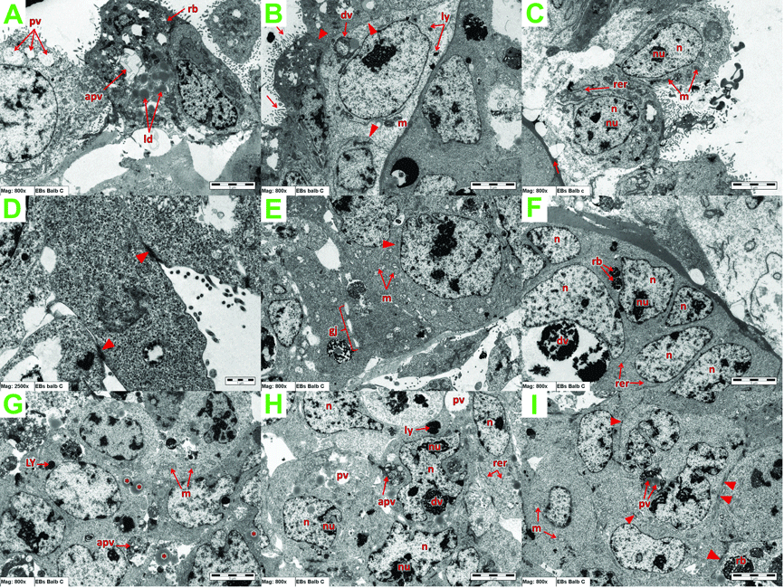
| Figure 4: TEM showing the cells in Balb/C EBs. dv: digestive vacuole, m: poorly developed mitochondria, ly: lysosomes, microvilli (arrows), tight junction (arrows head). (B) m: poorly developed mitochondria, microvilli (arrows), tight junction (arrows head). (C) n: nucleus, nu: nucleolus, m: well-developed mitochondria, rer: stalks of RER, basement-like membrane (arrow). (D) desmosomal like junction (arrows head). (E) tight junction (arrow head), m: well-developed mitochondria, gj: gab junction. (F) n: nucleus, nu: nucleolus, rer: stalk of RER, dv: digestive vacuole, rb: residual bodies.(G) m: well-developed mitochondria, lipid droplets (*), ly: lysosome, apv: autophagocytic vacuole. (H) n: nucleus, nu: nucleolus, rer: stalk of RER, pv: phagocytic vacuole, dv: digestive vacuole, ly: lysosome, apv: autophagocytic vacuole. (I) dismosomal like junction (arrows head), rb: residual bodies, m: well-developed mitochondria, ld: lipid droplets. |