|
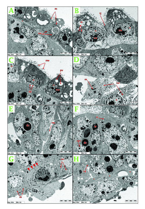
| Figure 6: TEM showing the terminal and peripheral cells in 129 W9.5 EBs. (A) m: juvenile mitochondria, rer: reticular stalk of rER, basment-like structure (*). (B) tj: tight junction, n: nucleus, nu: nucleolus, m: poorly developed mitochondria. (C) ld: lipid droplets, mv: microvilli, pv: phagocytic vacuole. (D) m: elongated mitochondria, rer: dilated rER. (E) dv: digestive vacuole, mv: microvilli. (F) m: well-developed mitochondria, ly: lysosomes, apv: autophagocytic vacuole. (G) m: poorly developed mitochondria, rer: stalk of rER, desmosomal like junction (arrows head). (H) ld: lipid droplets, lipid plaques (*). |