|
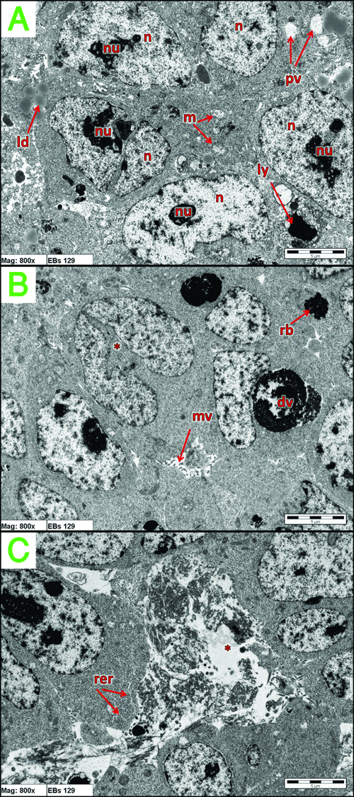
| Figure 7: TEM showing the middle cells in 129 W9.5 EBs. (A) n: nucleus, nu: nucleolus, pv: phagocytic vacuole, ly: lysosomes, ld: lipid droplets, m: welldeveloped mitochondria. (B) mv: microvilli, dv: digestive vacuole, rb: residual body, dividing cell (*). (C) rer: stalk of rough endoplasmic reticulum, indication of cell necrosis (*). |