|
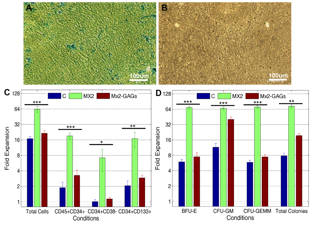
| Figure 1: Matrix modification and fold expansion of HSPCs for ablation experiment. (A-B) Alcian blue staining of untreated and GAGs ablated matrices respectively. Absence of bluish green staining in B indicates the ablation of GAGs after heparinise and chondroitinase treatment (scale bar = 100μm). (C) Represents the fold expansion of the HSPC subpopulation based on surface markers. (D) Represents the fold expansion of the CFUs compared to the fold expansion for total cells. Expansion of all HSPC lineages, except CFU-GMs, show significant reduction after GAGs ablation. Statistical analysis was done using One-way ANOVA (* p ≤ 0.05; ** p ≤ 0.01; *** p ≤ 0.005). Results are based on an average of three individual experiments with triplicates of each condition within an experiment. BFU-E (burst forming unit-erythroid), CFU-GM (Colony-forming unit Granulocyte, Macrophage) and CFU-GEMM (Colony-forming unit Granulocyte, Erythroid, Macrophage, Megakaryocyte). |