|
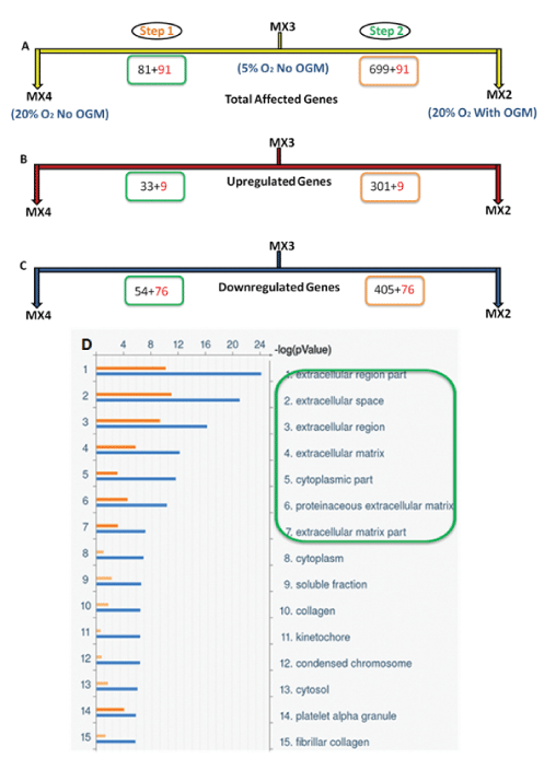
| Figure 2: Analysis of differentially expressed genes in step 1 and 2. The upper three panels show the graphical representation of the Venn diagram for differential genes. Step 1 represents MX3 vs MX4 and demonstrates ‘the effect of increase in O2’ whereas step 2 represents MX3 vs MX2 showing ‘the effect of both, increase in O2 and addition of osteogenic medium (OGM)’. (A) Indicates the number of total affected genes whereas (B and C) represent the number of up- and down- regulated genes respectively. The numbers in red color represents common genes between two steps, while that in black color represents number of genes unique to that step. (D) Indicates top 15 cellular localizations of the differentially expressed genes in step 1 and 2. The p value (log) is shown by orange color for step 1 and blue color for step 2. Green box highlights that 6 out of 7 top localizations are ECM-related; except “cytoplasmic part” which is fifth most significant localization affected. |