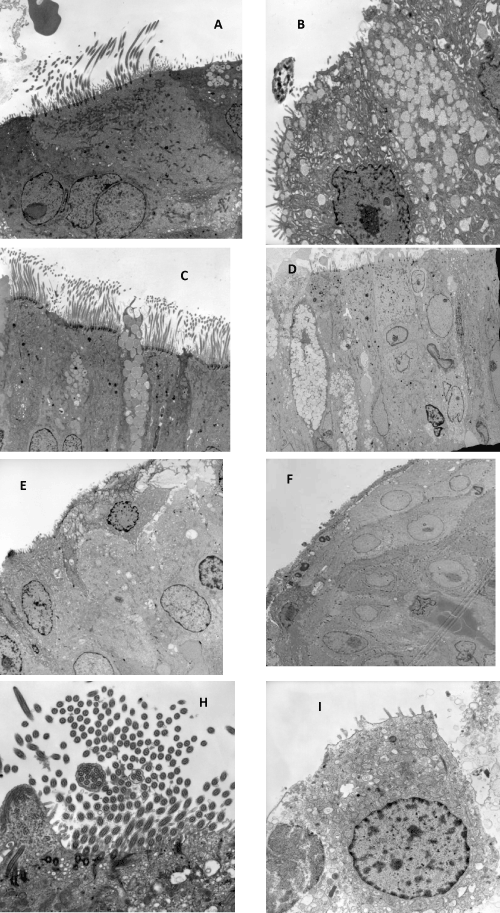
microscopy. (A) Ciliated cells with cilia decrease and microvilli increase. (B) Pseudostratified ciliated epithelium without cilia. (C) Normal ciliated
epithelium with goblet cells. (D) Ciliated epithelium without cilia and mononuclear cells migrating to the surface (arrow). (E) Epithelium into a process
of squamous metaplasia without cilia. (F) Squamous metaplasia. (G) Compound cilia. (H) Ciliated cells without cilia and mitochondria and with
cytoplasmic vacuolization.