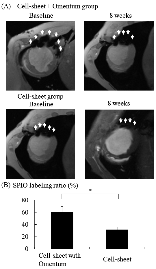
MRI, magnetic resonance imaging; SMB, skeletal myoblast. White arrow shows the low intensity of the super paramagnetic iron oxide-labeled area
MRI, magnetic resonance imaging; SMB, skeletal myoblast. White arrow shows the low intensity of the super paramagnetic iron oxide-labeled area |
| Figure 4: Cell tracking system by performing MRI. (A) MRI was examined just prior (baseline) and 8 weeks after SMB cell sheet implantation with or without omentum. (B) Cell retention was quantitatively assessed by performing MRI and the ironoxide-labeling method at 8 weeks corrected by that at day 0. There was significantly more cell retention of implanted SMB in the Cell sheet+Omentum group compared with in the Cell sheet group. |