|
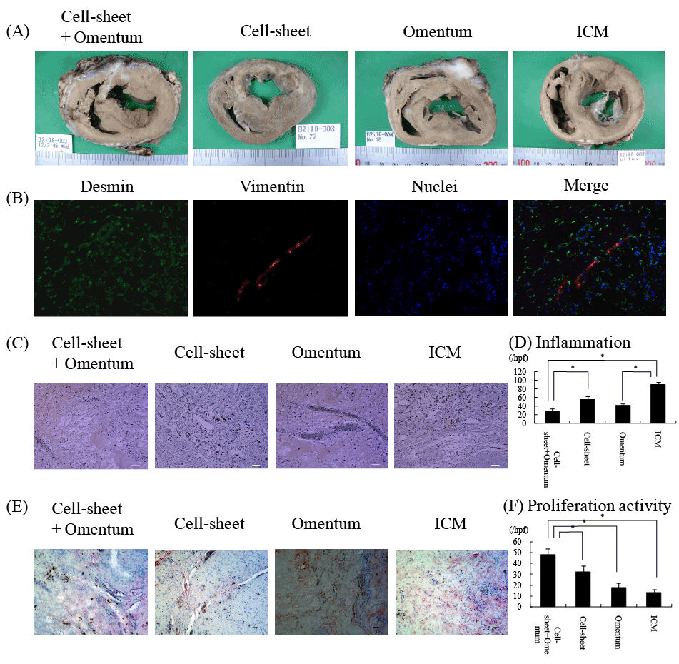
| Figure 5: Histological findings of extirpated hearts after treatment. (A) Macroscopic images of impaired myocardium after treatment. (B) Immunostaining fordesmin and vimentin showing desmin-positive andvimentin-negative cells in the implanted cell sheets following SMB cell sheet with omentum implantation. Green, desmin; red, vimentin; blue, nuclei. (C) Representative anti-CD68 staining of the border myocardium. (D) Quantification of inflammation. Inflammation at the border was significantly suppressed in the SMB Cell sheet+Omentum group compared with the SMB Cell sheet only group. (E) Representative anti-PCNA staining of the border myocardium. (F) Quantification of cell proliferative activity. Proliferative activity was significantly enhanced in the Cell sheet with Omentum group compared with the other groups. *p<0.05. SMB: skeletal myoblast; PCNA: proliferative cell nuclear antigen. |