|
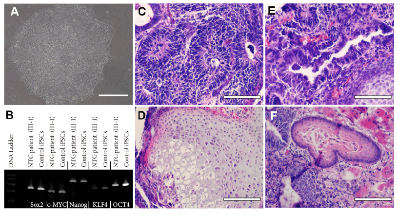
| Figure 2: Derivation of iPSCs from a patient affected with TBK1-associated Glaucoma. (A) Phase micrograph ofa TBK1-iPSC colony demonstrating a pluripotent morphology. (B) rt-PCR analysis of RNA isolated from WTiPSCs and TBK1-iPSCs targeted against pluripotency marker expression. (C-F) H&E staining of TBK1-iPSC derived teratomas that show each of the three embryonic germ layers ((C) ectoderm, (D) mesoderm, and (E and F) endoderm). Scale bar=100 microns. |