|
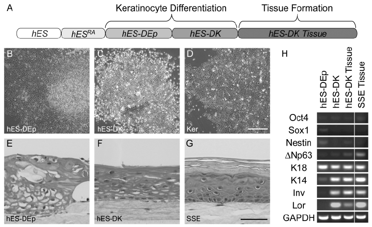
| Figure 2: hES-DK cells terminally differentiate and form keratinized stratified squamous epithelia. Schematic identifying each step of hESDK differentiation (A). hES cells treated with RA (hESRA) were harvested, maintained in suspension overnight, and passaged onto gelatin-coated plates in dKSFM. These cells (hES-DEp) were harvested prior to confluence and passaged once more onto gelatin-coated plates in dKSFM (hES-DK). Subconfluent hES-DK cells were harvested and seeded into organotypic culture for tissue formation. Representative phase contrast images of subconfluent hES-DEp (B), hES-DK (C), and epidermal keratinocytes (D). Histological analysis revealed the tissue architecture generated from organotypic culture of hES-DEp cells (E), hES-DK cells (F), and epidermal keratinocytes (G). RT-PCR detection of neural, epithelial, and keratinocyte terminal differentiation markers during hES-DK differentiation (H). Samples from hES-DEp and hES-DK cells, and the tissue generated from hES-DK cells, were evaluated. Results depict one of three independent differentiation series. Expression of Sox1 and nestin were confirmed by evaluating spontaneously differentiated hES cells. Stratified squamous epithelia (SSE) generated from epidermal keratinocytes were included as a comparison for own keratinocyte markers. Scale bar represents 500 µm (B,C,D); 50 µm (E,F,G). epidermal keratinocytes (Ker), keratin 18 (K18), keratin 14 (K14), involucrin (Inv), loricrin (Lor). |