|
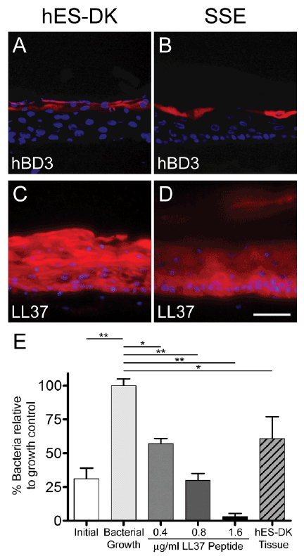
| Figure 5: Functional host defense peptides are expressed and appropriately localized in hES-DK tissue, as confirmed by antimicrobial activity in vitro against S. carnosus. Indirect immunofluorescent detection (red) of the host defense peptides hBD3 (A and B) and LL-37 (C and D) in stratified tissue generated from hES-DK and epidermal keratinocytes. Hoechst 33258 stain was used to visualize nuclei (blue). Results are representative of three independent experiments. Scale bar represents 50 µm. Extent of bacterial growth in the absence or presence of antimicrobial peptide or hES-DK tissue onditioned medium (E). Compared to the initial bacterial concentration, significant growth was observed in the bacterial growth control samples. The addition of LL37 peptide and conditioned medium significantly reduced the level of bacterial growth as compared to untreated control samples (*p<0.05; **p<0.01). Data represent mean ± standard deviation. |