|
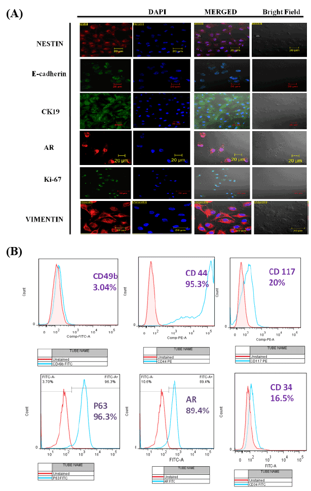
| Figure 2: Characterization of isolated prostate cells. (A) Immunofluorescence staining of BPH prostate cells for Nestin, E-cadherin, Ck19, Androgen Receptor, Ki-67 and Vimentin (first panel from left side). The second panel from left side shows DAPI staining and the second panel form right side shows the merged images. Bars represent 20 µm (B) FACS analysis of cell surface markers demonstrate 3.04% CD49b, 95.3% CD44, 20%CD117, 96.3% p63, 89.4%AR and 16.5% CD34 positive cells in isolated prostate cell |