|
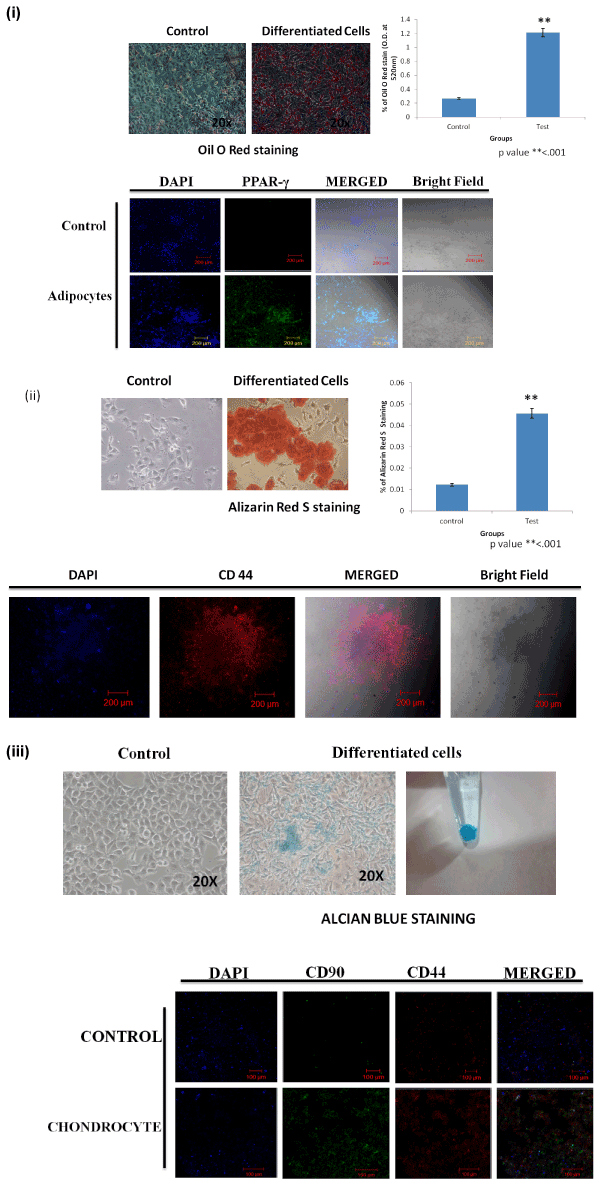
| Figure 5: Isolated prostate cells can differentiate to mesodermal cell lineages. (i) Oil Red O staining of isolated prostate cells after culture (A) without and (B) with adipocytic differentiation reagents (IBMax 10 mg/ml, 10 mM dexamethasone and 10 mg/l insulin) for 8 days. (A) Control cultures showed Oil Red O negative cells; (B) Positively staining adipocytes. (C) histogram showing the significant Oil Red O staining in differentiated cells. (D) Immunofluorescence staining of PPAR-γ for differentiated adipocytes. Nuclei were stained with DAPI. (ii) Alizarin red S staining of isolated prostate cells after culture (2A) without and (2B) with osteocyte differentiation reagents (20 mM glycerol phosphate, 50 µg/ml ascorbic acid, 10 mM dexamethasone and 10 mg/l insulin) for 10 days. (2A) Control cultures showed Alizarin red S negative cells; (2B) Positively staining osteocytes. (2C) histogram showing the significant Alizarin red S staining in differentiated cells. (2D) Immunofluorescence staining of CD44 for differentiated osteocytes. Nuclei were stained with DAPI. (iii) Alcian blue staining of isolated prostate cells after culture (A) without and (B) with chondrocyte differentiation reagents (10 mM dexamethasone and 10 mg/l insulin) for 20 days. (A) Control cultures showed Alcian blue negative cells; (B) positively staining chondrocytes. (C) Immunofluorescence staining of CD90 and CD44 for differentiated chondrocytes. Nuclei were stained with DAPI. |