|
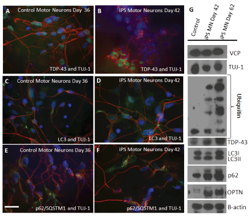
| Figure 4: Differentiation of human iPSC into neurons. Control and differentiated iPSC-derived neural lineage were double immunostained with (A and B) TDP-43 (FITC) and TUJ-1 (TRITC), (C and D) LC3 (FITC) and TUJ-1 (TRITC), and (E and F) p62/SQSTM1 (FITC) and TUJ-1 (TRITC) (Magnification 200X). DAPI was used for nuclear staining. (G) Western blot analysis of markers: VCP, TUJ-1, Ubiquitin, TDP-43, LC3-I/II, p62/SQSTM1, and Optineurin (OPTN). β-actin was used as a loading control in these experiments. |