|
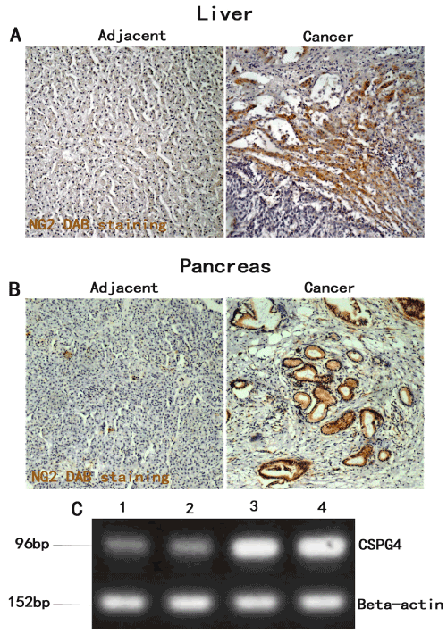
| Figure 1: Different Pattern of Expression of NG2/CSPG4 in Human Liver and Pancreatic Cancer Biopsies detected by 3,3′-diaminobenzidine (DAB) stainingand RT-qPCR. (A) DAB staining showed that highly positive staining of NG2 antigen in liver cancer, especially around blood vessels (right panel), while NG2 is negative or light positive in adjacent tissue of liver cancer (left panel). (B) Similar pattern showed in pancreatic cancer. (C) RT-qPCR showed highly expression of CSPG4 in metastatic tissues from liver (lane 3) and from pancreatic cancer patients (lane 4) compared to the adjacent liver tissue (lane 1) and the adjacent pancreatic tissue (lane 2) that all from the same patients. Original magnification, ×100. |