|
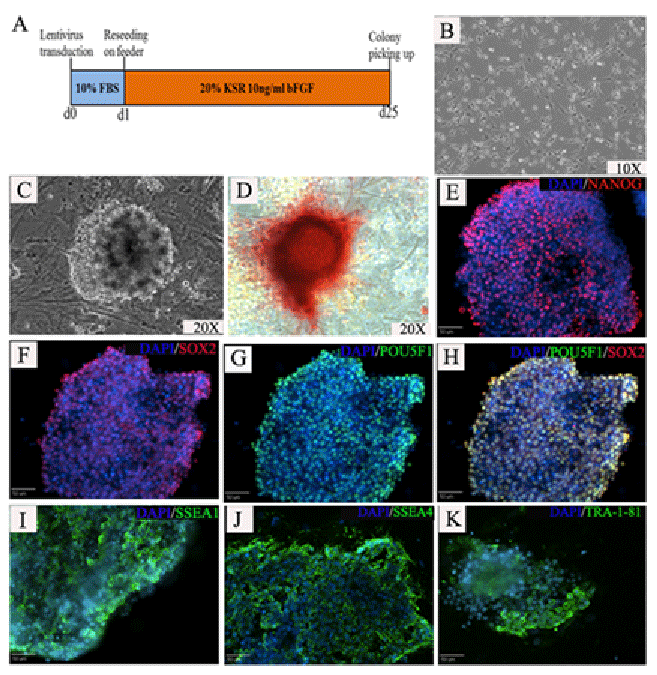
| Figure 1: Generation of piPSCs from pig fibroblast cells. Approximate time table of piPSCs generation (A). Pig fibroblast showed typical flattened morphology with extensions before transduction (B). Putative piPSCs grew as small dome shaped colonies showing well defined borders at day 15 post-transduction with single cells displaying large nucleoli and high nuclear to cytoplasm ratios typical of iPSC morphology (C). piPSCs stained positive for alkaline phosphatase (D). Immunostaining demonstrated that piPSCs were strongly positive for the pluripotent factors NANOG (E); Dapi nuclear marker shown in blue), SOX2 (F) and POU5F1 (G; H-POU5F1 and SOX2 merge). piPSCs were also positive for the stem cell specific surface antigens SSEA1 (I), SSEA4 (J) and TRA-1-81 (K). |