|
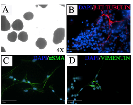
| Figure 5: Differentiation of piPSCs into cells representing all 3 germ layers. piPSCs formed compacted EBs (A) and underwent 10 days of EB differentiation. Immunostaining of plated EBs showed that cells were positive for the ectoderm marker βIII-TUB (B), mesoderm marker αSMA (C) and endoderm marker Vimentin (D). Scale bars=50um. |